WHAT IS CROSSLINKING?


An Effective Treatment for Keratoconus- and Possibly a Treatment to Reduce the Risk of Regression after LASIK or PRK.
Corneal collagen crosslinking is now the gold standard treatment for keratoconus, a progressive corneal degeneration. Corneal collagen crosslinking has been shown to stabilize the cornea in keratoconus, and stop the progression of this condition. Results of 5 years of follow up and international studies support the effectiveness and safety of this treatment of progressive corneal thinning. Because this procedure is not yet FDA approved in the United States, corneal crosslinking is available at King LASIK in Canada only.
More recently, crosslinking has been adopted by some surgeons for use in corneal infections, as well as an adjunct to primary laser vision correction procedures like PRK and LASIK, although the benefit of crosslinking in these settings remains controversial pending the outcome of clinical studies.
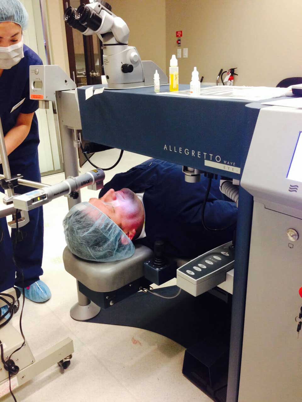
Corneal collagen crosslinking involves the application of riboflavin eye drops to the cornea, resulting in penetration of riboflavin into the corneal stroma, followed by exposure of the cornea to a measured dose of ultraviolet light irradiation. The UV light activates the riboflavin, resulting in enhanced crosslinking between the collagen fibers in the cornea. The enhanced crosslinking results in increased corneal strength and rigidity, with consequently arrested progression of steepening and often a significant amount of corneal flattening.
WHAT CAUSES KERATOCONUS?
Weak binding between the corneal collagen fibers is thought to be the underlying cause of keratoconus. Keratoconus may be a genetically linked condition. Due to weak binding between the corneal fibers the cornea may begin to stretch and bulge. Rubbing of the eyes may contribute to the worsening of keratoconus. Vision may begin to deteriorate as the irregular corneal shape results in astigmatism that progressively worsens due to the corneal bulging. The typical cornea in keratoconus is irregular shaped. A keratoconic cornea may have inferior steepening seen on corneal topographic mapping, corneal thinning seen on corneal thickness measurements known as pachymetry, and often increasing amounts of nearsightedness (myopia) and astigmatism. Eyeglasses or contact lenses may be needed to provide clear vision in the early stages of keratoconus.
In later stages, eyeglasses or contact lenses may not provide satisfactory visual acuity and a rigid contact lens may be needed. Traditionally, a full-thickness corneal transplant has been the definitive treatment for advanced keratoconus with unsatisfactory vision or contact lens intolerant patients. Full-thickness corneal grafts are not done as frequently now as newer techniques involving partial thickness corneal grafts (for example, Deep Anterior Lamellar Keratoplasty) are now excellent options to treat end-stage keratoconus.
HISTORY OF CROSSLINKING
Corneal collagen crosslinking has truly revolutionized the initial treatment of mild to moderate keratoconus that is not yet requiring a corneal transplant. In many cases, crosslinking will greatly reduce or eliminate the need for a corneal transplant if the crosslinking is performed at an earlier stage when it is most effective.
The original protocol for performing crosslinking was developed by Dr. Theo Seiler of Germany and it is referred to as the Dresden protocol. Because this technique has been used for the longest period of time, most of the long-term data that is being reported today are based on crosslinking performed using this protocol. When this therapy was first developed, it was used for patients who experienced progressive keratoconus. The rationale was that this new technology should be used for patients who probably will need a corneal transplant, and this new technology involves only a low risk to a patient who has no other non-surgical therapeutic options. At first, slowly progressive keratoconic patients were watched, avoiding unnecessary complications as indications for this new technology evolved. However, over time, the relative safety of corneal collagen crosslinking has been demonstrated. Now early diagnosis and treatment of keratoconus with corneal crosslinking is believed to prevent the rapid progression of this disease in all patients, including children and adolescents. If one waits for the progression of keratoconus, it may be too late, as there are fewer corneal collagen fibers to crosslink as the cornea thins and the vision deteriorates. Earlier treatment of keratoconus may arrest progression. Studies by Dr. Hafezi (Switzerland), and other European investigators such as Dr. Vinciguerra and Dr. Caporossi have shown that treatment of children and adolescents with keratoconus may be beneficial, as these younger patients will experience the same corneal flattening and arrest of keratoconus that is seen in the adult population. As a result of these studies, a general recommendation was made at the 9th International Corneal Crosslinking Congress in Dublin, Ireland in December 2013 that children and adolescents should be treated as soon as the diagnosis is made, without waiting for progression.
THE FUTURE OF CORNEAL COLLAGEN CROSSLINKING
New trends in corneal collagen include the consideration of epithelial on crosslinking versus the traditional epithelial off crosslinking. Epithelial off crosslinking, otherwise known as transepithelial corneal collagen crosslinking, is similar to the traditional crosslinking technique, except that the corneal epithelium is not removed. Riboflavin, when not combined with dextran (typically used in epithelial off crosslinking), may penetrate into the corneal stroma. However, the penetration of riboflavin through intact corneal epithelium may be limited and the efficacy of the procedure may be limited as a result. This is an area of ongoing clinical research. In addition, the use of a greater concentration of UV light during corneal crosslinking may allow a shorter treatment time. The traditional energy fluence with corneal collagen crosslinking has been 3 mW/square centimeter for 30 minutes, although newer accelerated protocols involve the application of UV light at 9mW/square cm for 10 minutes, or 30mW/square centimeter for as little as 3 minutes. The relative effectiveness of transepithelial crosslinking and higher energy irradiation remains unknown.
David Tonboul, MD of Bordeaux, France has shown with corneal confocal microscopy that corneal cells called keratocytes are temporarily lost following corneal collagen crosslinking. In addition, there is a loss of the corneal nerve plexus, but no effect on the corneal endothelium, the layer of corneal cells that pumps fluid from the cornea. It is unknown whether corneal collagen crosslinking is effective without damage to corneal keratocytes, and what role the corneal keratocytes and corneal nerve plexus have in determining the effectiveness of corneal collagen crosslinking. Dr. Tonboul hypothesizes that keratocyte loss is an important measure of the effectiveness of corneal collagen crosslinking. In his study that compared transepithelial crosslinking to epithelial off crosslinking, there was a very large loss of keratocytes in the epi off the group, but the transepithelial group was similar to the control eyes that received no treatment at all. The lack of penetration of riboflavin through intact corneal epithelium may be the reason.
LASIK X-TRA
The combination of LASIK and crosslinking, sometimes referred to as LASIK X-tra is controversial. At this time, there is no evidence to justify the combined use of corneal collagen crosslinking for every routine LASIK procedure to reduce the risk of corneal ectasia because the risk of corneal ectasia or thinning after routine LASIK in normal patients is so low. In addition, it is not known what effect crosslinking may have on LASIK outcomes, and when a LASIK retreatment should be offered, because the effects of crosslinking may evolve over months to years. Some doctors, such as Dr. Kannelopoulos of Greece have advocated LASIK X-tra for hyperopic patients, and for myopic patients who meet certain criteria that may increase the risk of regression: a refractive error greater than -6 diopters; age under 30 years; astigmatism greater than 1.5 diopters; or an astigmatism difference between eyes greater than 1 diopter. His technique involves routine LASIK followed by the application of 0.10% saline diluted riboflavin to the stromal LASIK bed. Following a 60 second soaking, the flap is repositioned, the interface is irrigated and the cornea is irradiated for 60-80 seconds at 30mW/square centimeter. Typically, this technique adds to the cost of the LASIK procedure, and the longterm effects and benefits are not clearly established. Crosslinking could be performed after LASIK or PRK, if needed or if future studies were to show a benefit.
As this discussion illustrates, corneal collagen crosslinking techniques, technology, and indications are evolving. We know that crosslinking is highly effective for treating keratoconus. The most effective protocol and all applications for this technology remain unknown at this time.
At King LASIK, after careful consideration of the patient’s clinical status, preference, and consultation, Dr. King may offer either a patient either epithelial on or epithelial off corneal collagen crosslinking. We use both the traditional Dresden protocol and accelerated corneal crosslinking with higher light energy. Refractive surgery, usually PRK in combination with crosslinking is offered to select patients. If you have keratoconus, your previous records are an essential part of your evaluation and we would request that your records are available at the time of your consultation. Please be advised that this information presented may be subject to change as clinical studies may support the refinements of crosslinking techniques and technology.
Please contact King LASIK in Canada for further information or to schedule a consultation with Dr. King.
